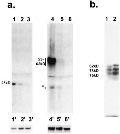
FIG. 9.
Phosphorylation of IE62 peptides in transfected cells by the ORF66 protein kinase. (a) Phosphorylated peptides were immunoprecipitated from cells transfected with plasmid pGK2-HA5 (lanes 1 to 3) or from pGK2-HA17 (lanes 4 to 6) following cotransfection with pKCMV66 (lanes 1 and 4), pGK2-HA66 (lanes 2 and 5), or pKCMV66ct2 (lanes 3 and 6). Proteins were immunoprecipitated with a mixture of polyclonal and monoclonal antibodies to the IE62 protein. The approximate sizes of the main phosphorylated species in kilodaltons are indicated at the left. The asterisk indicates a suspected breakdown product of the 55- to 62-kDa product. The 55- to 62-kDa species detected in lanes 4 to 6 migrate as a smear due to the close proximity of the heavy chain of IgG in the immunoprecipitates. Lanes 1′ to 6′ represent the main species of proteins detected by immunoblotting with anti-IE62 antibodies in whole-cell extracts from a parallel transfection carried out without the presence of radiolabel. (b) Phosphorylated peptides expressed from cells transfected with pKCMV62d1-587, either with (lane 2) or without (lane 1) pKCMV66 at an equimolar amount. The approximate sizes of the species detected in cells transfected without the ORF66 protein kinase are shown in kilodaltons.

