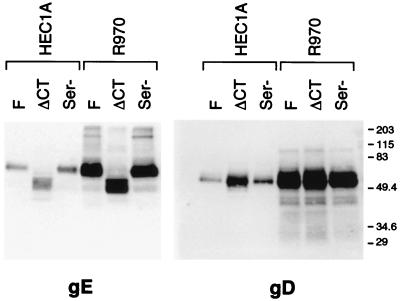
FIG. 7.
Incorporation of gE and gD into virions. HEC-1A epithelial cells or R-970 cells were infected with wild-type HSV-1 (strain F), F-gEΔCT, or F-gESer, and then cell culture supernatants from the cells were collected at 22 h, before the cells had rounded. Virus particles were collected from cell culture supernatants and centrifuged through a cushion of 30% sucrose, and detergent extracts of the virions were subjected to electrophoresis in polyacrylamide gels. Blots were incubated with anti-gE MAb 3114 or anti-gD MAb DL6, washed, incubated with horseradish peroxidase-conjugated goat anti-mouse IgG, and washed again, and proteins were visualized using enhanced chemiluminescence. The positions of molecular mass markers are shown along the right side of the gel.

