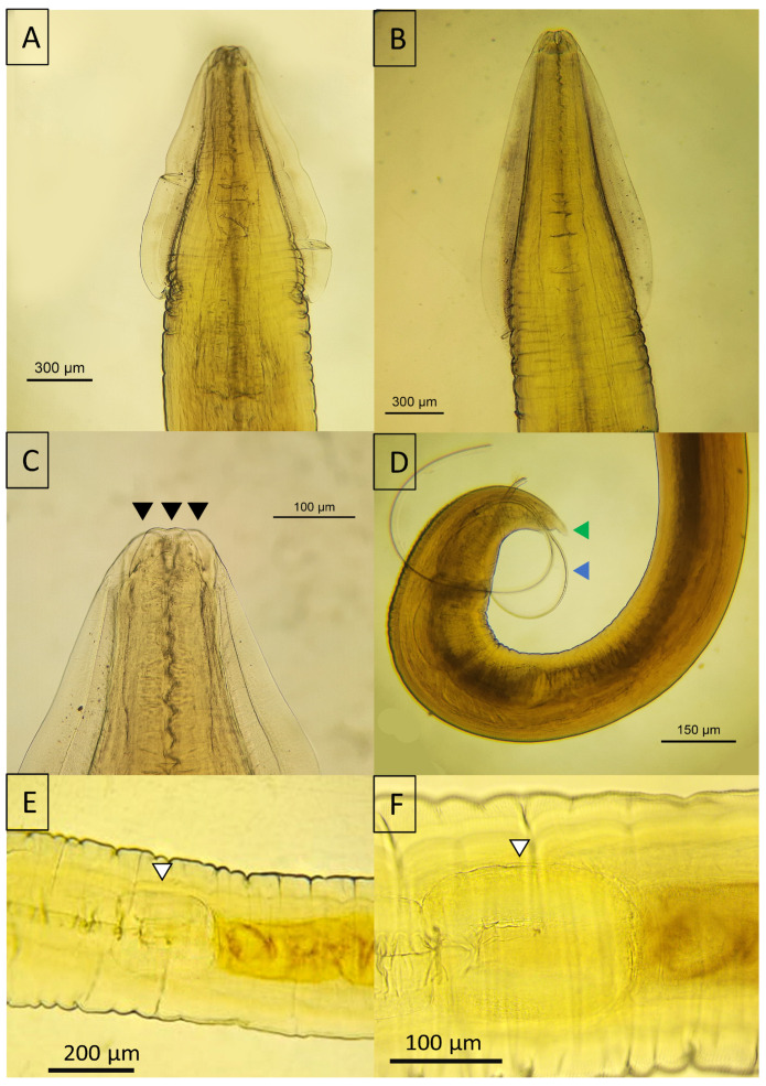
Figure 7.
Micrographs of Toxocara spp. detected in Panthera onca. (A) Cervical wing of Toxocara cati; (B) cervical wing of Toxocara canis; (C) anterior view of T. cati showing the three lips; (D) posterior view of a male T. canis with both spicules externalized. (E,F) Ventriculus that intercalated between the esophagus and the intestine in T. canis. Black arrowhead—lips; blue arrowhead—spicules; green arrowhead—digitiform process; white arrowhead—ventriculus.

