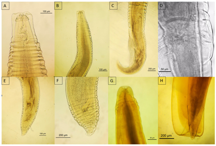
Figure 9.
Micrograph of Physaloptera spp. recovered from wild felids. (A,B) Anterior portion of Physaloptera anomala from Leopardus pardalis; (C) posterior portion of male P. anomala; (D) showing the sessile papillae; (E) posterior portion of female P. anomala; (F) posterior end, part of the uterus filled with eggs; (G) anterior portion; (H) posterior portion of female showing the cuticular sheath of Physaloptera praeputialis in Herpailurus yagouarandi.

