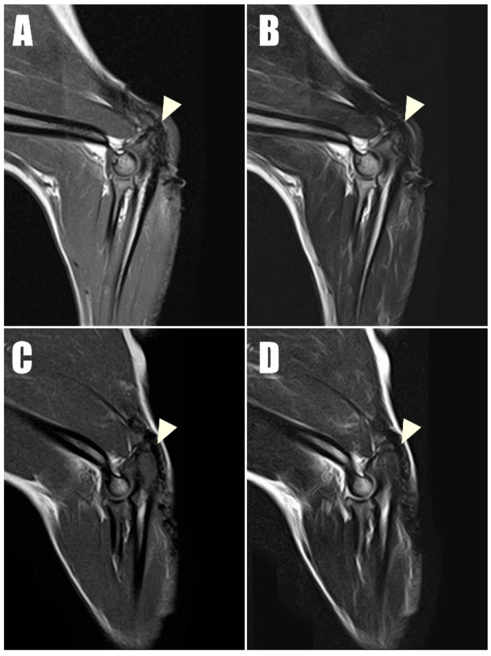
Figure 8.
Postoperative magnetic resonance imaging (MRI) taken one year postoperatively captured (A) T1-weighted and (B) T2-weighted sagittal images of the right triceps brachii tendon, along with (C) T1- T1-weighted and (D) T2-weighted sagittal images of the left of triceps brachii tendon. These images demonstrate low signal intensity on both T1 and T2 sequences, indicating intact connectivity of the tendon.

