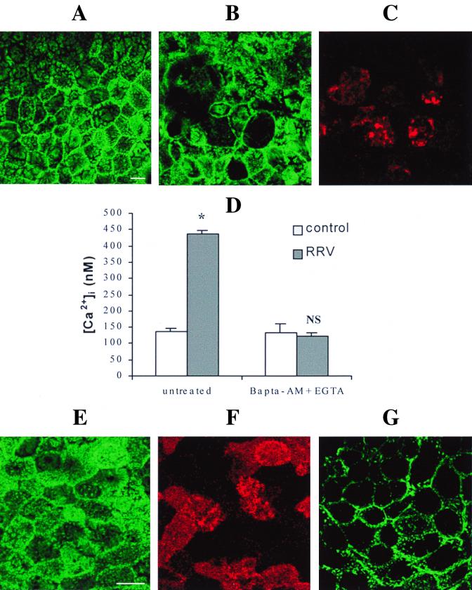
FIG. 2.
Microvillar F-actin disassembly induced by RRV infection (A to C). At 18 h p.i., RRV-infected (B) and mock-infected (A) Caco-2 cells were fixed, permeabilized, and stained with fluorescein-phalloidin, which binds to F-actin. RRV proteins were immunostained with polyclonal anti-group A rotavirus antibody and rhodamine-labeled anti-rabbit immunoglobulin G antibody. A horizontal section was generated by CLSM at the apex of the cells. (C) RRV staining in the same cells as in panel B. Treatment of infected cells with EGTA and BAPTA-AM totally abolishes the RRV-induced [Ca2+]i rise (D). Infected Caco-2 cells were treated at 15 h p.i. for 3 h with 3 mM EGTA and 75 μM BAPTA-AM. At 18 h p.i., cells were trypsinized to measure [Ca2+]i by quin2 fluorescence. Values are means ± standard deviations from three experiments. Statistical differences between control and infected cells were determined by Student's t test. NS, not significantly different; ∗, P < 0.01. EGTA and BAPTA-AM treatment totally protects Caco-2 cells from RRV-induced microvillar F-actin alteration (E and F). (E) Microvillar F-actin staining in treated 18 h p.i.-infected cells; (F) RRV staining in the same cells as in panel E; (G) microvillar F-actin disorganization in Caco-2 cells stained with fluorescein-phalloidin after treatment for 7 min with 10 μM ionomycin. Bars, 10 μm.

