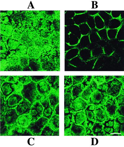
FIG. 8.
Effect of supernatants of RRV-infected Caco-2 cells on microvillar F-actin organization of uninfected Caco-2 cells. At indicated times after the addition of 18 h p.i.-infected or mock-infected cell supernatants, Caco-2 cells were fixed, permeabilized, and stained with fluorescein-phalloidin. A horizontal section was generated by CLSM at the apex of the cells. F-actin staining 40 min after the addition of supernatants of mock-infected cells (A) or infected cells (B). (C) Normal F-actin pattern 90 min after the addition of supernatants of infected cells. Treatment of Caco-2 cells with 3 mM EGTA and 50 μM tBuBHQ (Fig. 7) prevented microvillar F-actin disorganization induced by supernatants of 18 h p.i.-infected cells (D). Bar, 10 μm.

