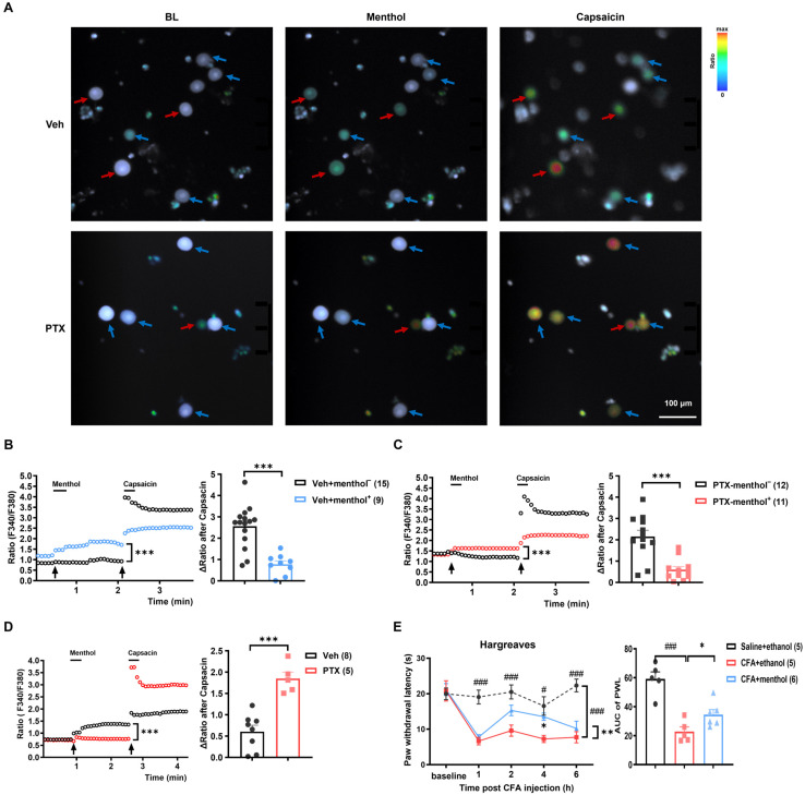
Figure 7.
Activation of TRPM8 by menthol inhibited the function of TRPV1 in vitro and in vivo. (A–D) The calcium imaging experiment demonstrated that the DRG neurons pre-activated by 200 μM menthol showed a significantly decreased response to 1 μM capsaicin. (A) Representative calcium images of DRG neurons from PTX-treated and vehicle-treated rats. Neurons responsive to menthol are indicated by red arrows (menthol+), while those unresponsive to menthol are indicated by blue arrows (menthol−). (B) Statistical analysis of the calcium signal changes after capsaicin treatment (ΔRatio after capsaicin) in the menthol-responsive and unresponsive neurons from the vehicle-treated rats. (C) Data analysis of calcium signal changes in DRG neurons in response to capsaicin between the menthol-responsive and unresponsive groups from the PTX-treated rats. (D) Comparison of the overall (including both menthol+ and menthol− neurons) calcium signal changes to capsaicin in the vehicle and PTX groups after activation of TRPM8 by menthol. Data were analyzed using a two-way ANOVA followed by Bonferroni post hoc tests, 3 independent experiments, * p < 0.05, *** p < 0.001, n = 15, 9 in (B); n = 12, 11 in (C); n = 8, 5 in (D). (E) The topical application of 1% menthol significantly attenuated CFA-induced inflammatory heat hyperalgesia. Left: Paw withdrawal latency (PWL) in Hargreaves test for control, CFA, and CFA/menthol groups. Right: Statistical analysis of PWL from the left figure. Black arrows in the left panels of (B–D) indicate the time point of administration of menthol or capsaicin. All data are presented as means ± SEM. Data were analyzed using a two-way ANOVA followed by Bonferroni post hoc tests and an unpaired t-test. * p < 0.05, ** p < 0.01, *** p < 0.001, # p < 0.05, ### p < 0.001, n = 5, 5, 6.

