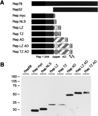
FIG. 1.
Rep proteins and chimeric proteins generated to study DNA binding. (A) Schematic of wild-type and chimeric Rep proteins. The amino-terminal 244 residues of Rep78 were tagged with a Myc epitope, joined to the SV40 large T NLS, and fused to the LZ or a modified zipper (TZ) of GCN4. The transcriptional activation domain of VP16 (AD) was included in some fusion proteins. Drawings are not to scale. (B) Western blot analysis of chimeric Rep proteins. Nuclear extracts were made from 293 cells transfected with plasmids expressing the indicated fusion proteins. Proteins were separated on a 10% polyacrylamide gel by SDS-PAGE, and chimeric proteins were detected with an anti-Myc antibody. The positions of molecular size markers are indicated on the left.

