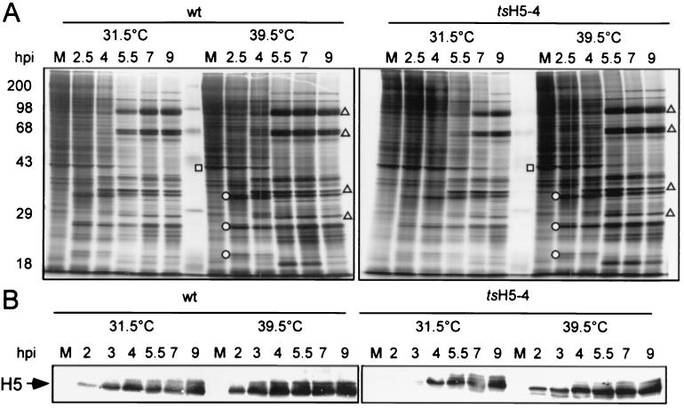
FIG. 4.
Protein expression in tsH5-4-infected cells. (A) BSC40 cells were infected with wt or tsH5-4 virus at an MOI of 15 and maintained at 31.5 or 39.5°C. The cells were metabolically labeled with [35S]methionine for 45 min before being harvested at the indicated time points (2.5, 4, 5.5, 7, and 9 h p.i.). As a control, uninfected cells were labeled with [35S]methionine for 45 min and analyzed in parallel (lane M). Cells were harvested and analyzed directly by SDS-PAGE on a 12% polyacrylamide gel. After electrophoresis, the gel was dried and exposed for autoradiography. The circles indicate early viral proteins, and the triangles indicate late viral proteins. The square denotes a host protein whose production is shut off after infection. (B) H5 protein accumulation. Samples prepared as above were analyzed by SDS-PAGE, transferred to nitrocellulose, probed with a polyclonal anti-H5 antiserum, and detected using an alkaline phosphate-conjugated secondary antibody. Only the relevant portion of the blot is shown.

