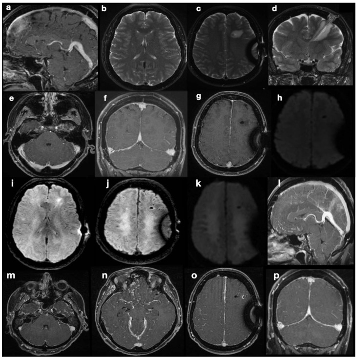
Figure 2.
Post-shunting MRI scans. The first post-operative MRI (a–h) is highly suggestive of intracranial hypotension, with “slumping midbrain” and depression of the vena magna Galeni (a), small ventricles (b), and supra- and sub-tentorial dural thickening with turgidity of the venous sinuses (f–h). At the same time, a fluid effusion surrounds the parenchymal course of the catheter, hinting at draining dysfunction. DWI sequences did not show abnormal findings (e). MR scan after marked neurological deterioration (i–p) shows ongoing demyelination of deep periventricular white matter in both hemispheres (i–k). Radiological signs suggestive of intra-cranial hypotension are even more conspicuous here than previously described in images (a–h).

