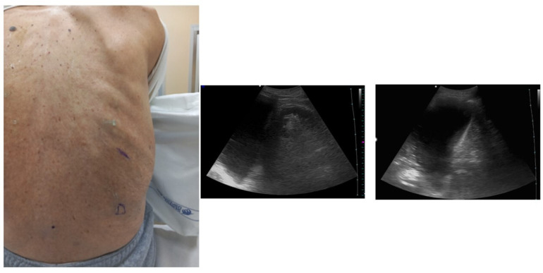
Figure 1.
Position of the patient with posterior approach and identification of the landmarks. Ultrasound allows one to identify two landmarks: the apex of the diaphragm at the end of expiration (marked D in the image) and cranially the point (intercostal space) chosen for insertion of the needle. Ultrasound allows one to optimally identify the point where the procedure should be performed. The choice is based on the visualization of the liquid as an anechoic space. The point where the presence of anechoic fluid will be most evident should be the optimal one to perform the procedure. Often at this point the effusion takes on a “V” morphology, delimited on the sides by the diaphragm and the atelectatic lung. The apex of the “V” would represent the specific point where the needle should be inserted and directed.

