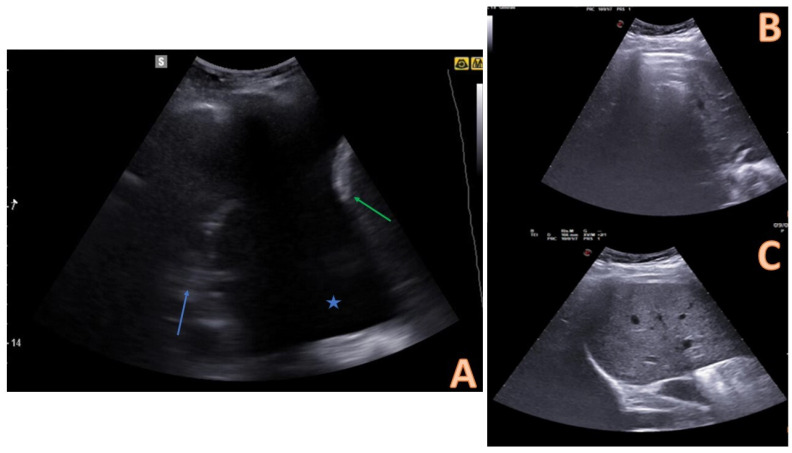
Figure 2.
Comparison between pleural effusion (A) and healthy lung on ultrasound (B,C). In (A), the pleural effusion is visible below the chest wall and ribs as an anechoic space (blue star). The effusion is delimited inferiorly by a hyperechoic formation that represents the diaphragm (green arrow), and superiorly by an echogenic structure that identifies the atelectatic lung parenchyma (blue arrow). (B,C) represent the appearance of the healthy lung base on ultrasound: an air interface with A (horizontal) lines is evident. This artefactual image covers the abdominal organ in inspirium (B) and uncovers it in expirium (B). This sign is called a “curtain sign.” When a pleural effusion is present, the curtain sign cannot be observed.

