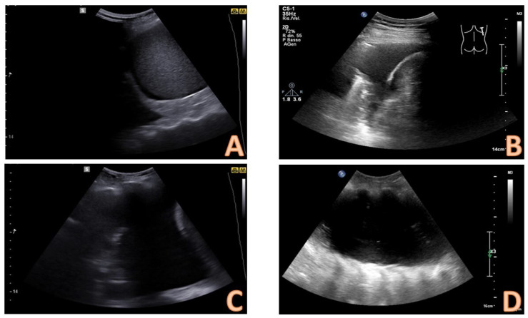
Figure 3.
Quantitative evaluation of pleural effusion. This assessment should preferably be performed with the patient seated. It is possible to provide a semi-quantitative estimate of the effusion by counting the number of intercostal spaces occupied by the effusion through longitudinal scans starting from the diaphragm and going up cranially. In image (A), there is a minimal effusion, visible only in the costophrenic sinus; in image (B), the effusion occupies only an intercostal space and is therefore of a mild degree; in image (C). the effusion extends for 2 intercostal spaces and is therefore of moderate degree; in image (D), the entire scan is occupied by the effusion with an extension > 3 intercostal spaces and is therefore massive.

