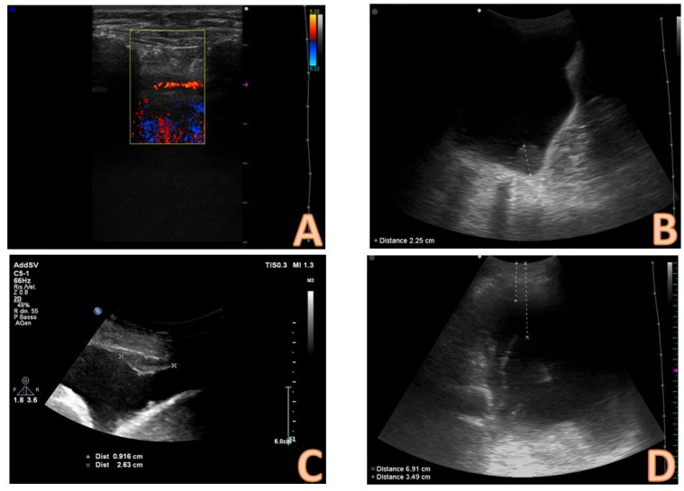
Figure 5.
Focused ultrasound evaluation of the needle insertion point. In image (A), an anomalous course of an arterial vessel in the intercostal space is evident; therefore, there is a high risk that the needle could injure the vessel, and this finding must induce the operator to vary the choice of insertion point. In image (C), a pleural plaque is evident; to insert the needle into the point where this plaque is present can both induce difficulties in inserting the needle and increasing the risk of bleeding from the plaque itself; the needle insertion point should be changed. Image (B) demonstrates how a pre-procedural ultrasound evaluation can highlight solid masses at the pleural level; this finding raises the suspicion of a malignant nature of the effusion. In image (D), there is an example of the evaluation of the distances between skin and effusion and between skin and lung, which can be done by ultrasound; this evaluation guides the choice of the needle and its insertion depth.

