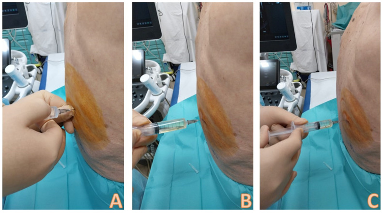
Figure 7.
Local anesthesia with lidocaine 1–2%. An ampule of lidocaine is drawn into a 10 mL syringe. In (A), the needle insertion phase is visible; the injection is performed at the chosen point and intercostal space. The non-dominant hand palpates the lower rib of the intercostal space; the dominant hand inserts the needle just above the upper edge of the lower rib; the needle is always directed downwards and never upwards to avoid damaging the intercostal artery that runs along the lower edge of the upper rib. The needle must pass the pleura and enter the pleural cavity, so that the local anesthetic can act on the richly innervated pleura; this is demonstrated by the fact that pleural fluid is aspirated into the syringe (B). At this point, boluses of 1–2 cc of anesthetic are injected at different points following a backward way to the subcutaneous tissue (C).

