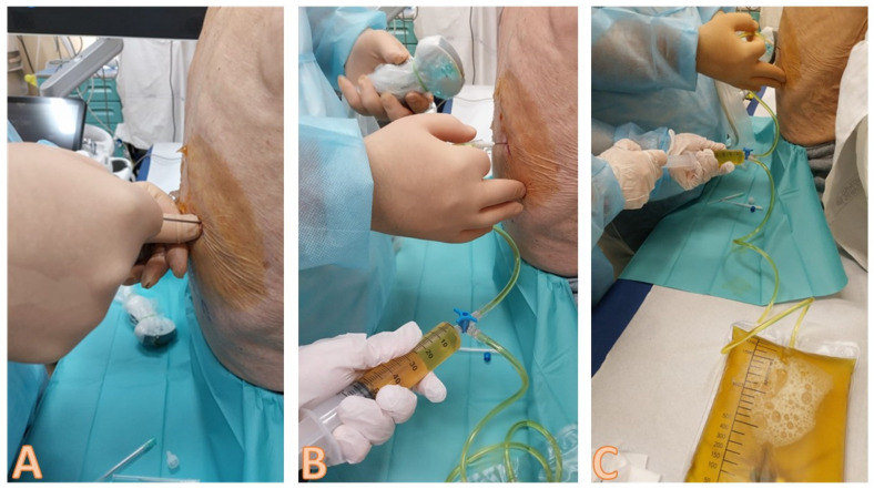
Figure 8.
Sequence of images about the insertion of the needle and the execution of the thoracentesis. In image (A), the non-dominant hand palpates the lower rib of the chosen intercostal space, and the dominant hand inserts the needle above the upper edge of the rib, always directing it downwards; it should be noted that, as it is an ultrasound-assisted procedure, the probe is not held in the hand when the needle is inserted, but is available to the operator in the sterile field with a sterile probe cover. In (B,C), the leakage of the pleural fluid through the collection system is highlighted, with a second operator who sequentially aspirates and allows the fluid to flow into the collection bag. Note how the ultrasound probe is taken back into the hand and used to verify the position of the needle and the residual amount of pleural effusion.

