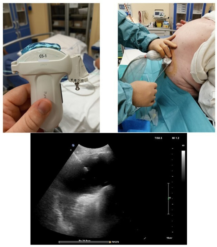
Figure 10.
Ultrasound-guided thoracentesis. In this example, we used a guide coupled on a convex probe, which creates a pre-established needle insertion angle. An oblique intercostal scan that allows one to better highlight the pleura and the effusion is performed. The needle is displayed along its entire path, and the exact position of the tip can be observed for the entire duration of the procedure.

