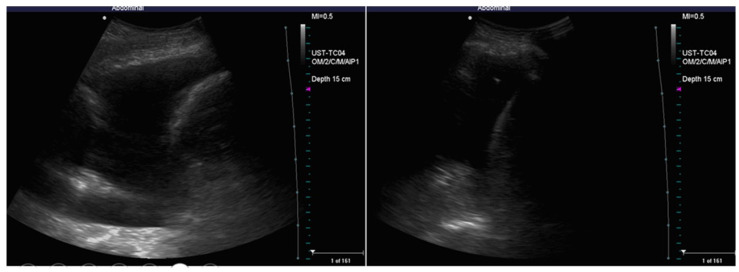
Figure 11.
Sequential ultrasound check of the effusion and the position of the needle during thoracentesis. There is a progressive reduction of the anechoic space with progressive re-expansion of the atelectatic lung with the appearance of internal air bronchograms. When the tip of the needle is too close to the re-expanded lung (right figure), the procedure should be stopped.

