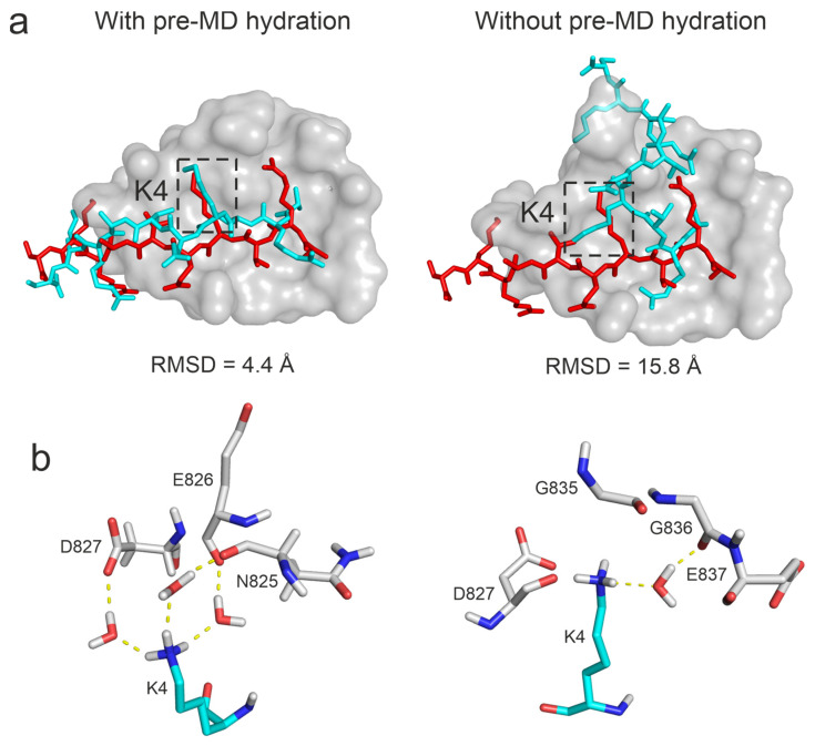
Figure 6.
Comparison of ligand binding modes produced with and without the pre-MD hydration step (Protocol P4, System 3o33). Target, MD-refined, and experimental reference ligand structures are depicted as grey surface, cyan, and red sticks, respectively (a). The close-up of the surrounding of ligand residue K4 (b), marked by dashed boxes at the top. Three bridging water molecules can be observed if pre-MD hydration was applied, while only one water bridge was formed at a wrongly found pocket without pre-MD hydration (b).

