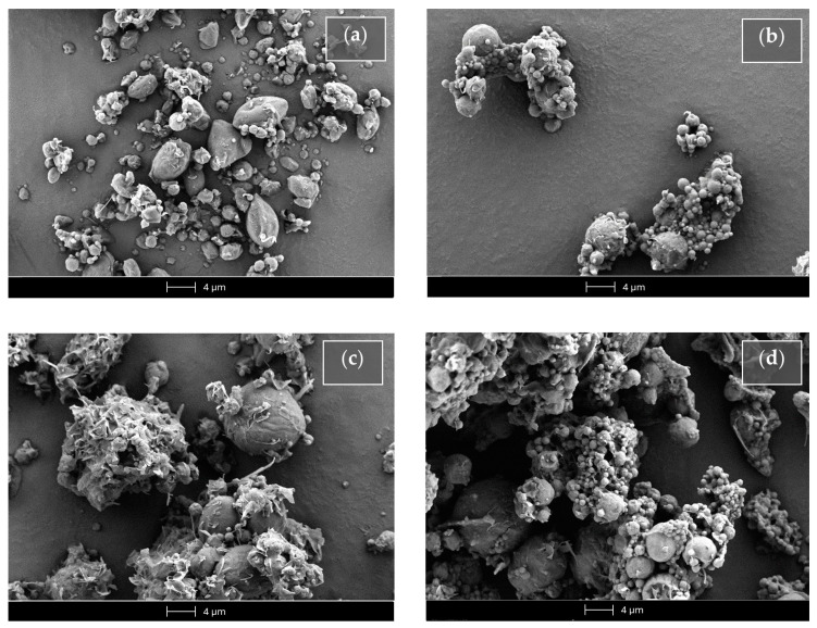
Figure 10.
Scanning electron microscopy (SEM) micrographs of the SLMs based on tristearin in the absence (Ts SLMs) or in the presence (Ts-glu SLMs) of glucose. (a) Unloaded tristearin-based SLMs. (b) Tristearin-based SLMs loaded with Fer-Ger. (c) Unloaded tristearin-based SLMs in the presence of glucose. (d) Tristearin-based SLMs in the presence of glucose loaded with Fer-Ger.

