Figure 4.
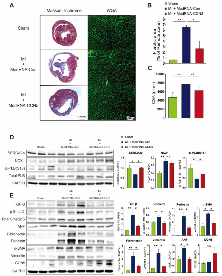
Therapeutic intervention with ModRNA-CCN5 reduces MI-induced cardiac dysfunction and fibrosis. (A) The heart tissues from the therapeutic intervention were used for all experiments. The red-stained area indicated normal tissue and the blue-stained area indicated fibrotic tissue in Masson’s trichrome staining. WGA staining was performed to measure the cellular size of the heart. The green-stained area indicated a cellular membrane. Scale bar: 50 μm. (B) Fibrotic area was analyzed using a computer-assisted method. ImageJ plug-in software was used to measure the ratio of fibrotic area. (C) Cell surface area (CSA) was analyzed using a computer-assisted method. ImageJ plug-in software was used to measure CSA. (D) Cardiac tissue lysates were immunoblotted with antibodies against SERCA2a, NCX1, p-PLB (Ser 16), total-PLB, and GAPDH. (E) Additional lysates were immunoblotted with antibodies against TGF-β, p-Smad2, Smad2/3, ANF, Fibronectin, Periostin, α-SMA, Vimentin, CCN5, and GAPDH. For each experimental group, animals were used as follows: n = 7 for sham, n = 7 for ModRNA-Con, and n = 8 for ModRNA-CCN5, * p < 0.05, ** p < 0.01, n.s.: not significant.

