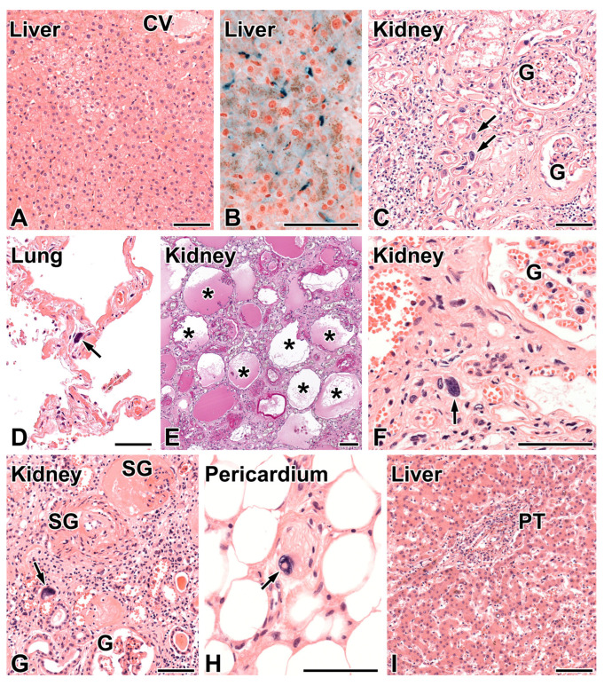
Figure 1.
Morphologic findings in the FAN1-mutation related disease. (A) Patient C, liver biopsy. Focal, non-specific degeneration of hepatocytes in the intermediary zone of liver acinus. CV—central vein. Hematoxylin and eosin (HE), 20×. (B) The Prussian blue staining demonstrated hemosiderin (blue) located exclusively in Kupffer cells, 40×. (C) Patient A, end-stage kidney. Karyomegaly of tubular epithelial cells (arrows), tubular atrophy, and interstitial fibrosis with focal and mild lymphocytic infiltrates can be seen. G—patent glomerulus. HE, 20×. (D) Patient A, lung. Karyomegaly in a non-specified cell of alveolar septum (arrow). HE, 20×. (E) Patient B, end-stage kidney. The widespread loss of tubules and cystic dilation of atubular glomeruli (asterisks) can be seen. Periodic acid–Schiff, 10×. (F) Patient B, end-stage kidney. Bizarre karyomegaly in a tubular epithelial cell (arrow) is present. G—patent glomerulus. HE, 40×. (G) Patient E, end-stage kidney. Karyomegaly of tubular epithelial cell (arrow). G—patent glomerulus, SG—sclerosed glomerulus. HE, 20×. (H) Patient E, subepicardial tissue. Bizarre karyomegaly in the Schwann cell (arrow) of a small peripheral nerve. HE, 40×. (I) Patient E, liver. The hepatocytes and sinusoids do not show specific abnormality. The portal tract (PT) shows mild infiltration mainly by lymphocytes. “Onion-skin fibrosis” of the bile duct is not present. HE, 10×. The bar represents 100 μm.

