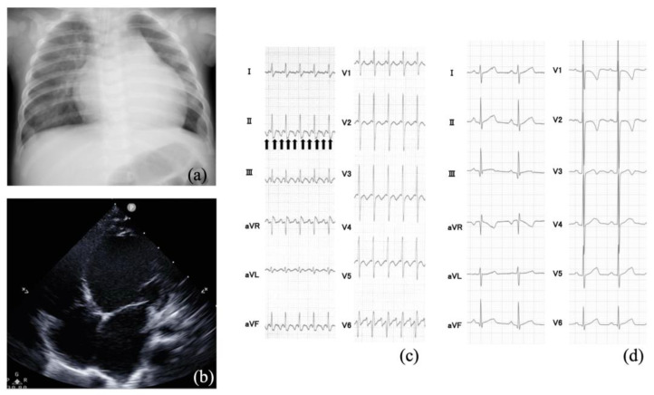
Figure 2.
(a) Chest X-ray showing cardiomegaly with a cardiothoracic ratio of 0.62 and lung congestion. (b) Echocardiography shows that the left ventricular dilation and wall motion are diffusely decreased. (c) Typical atrial flutter with 2:1 conduction. The arrows indicate the flutter waves. (d) Sinus rhythm after synchronized electrical cardioversion.

