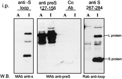
FIG. 3.
The loop region between TM1 and TM2 is not cytoplasmically exposed and accessible to specific antibody in microsomal membranes. Microsomes from DHBV-positive primary duck hepatocytes were incubated with antiserum to either the first loop region in the S domain (aa 207 to 226), the pre-S domain (aa 127 to 156), or the second loop region in the S domain (aa 267 to 284) or with a control antibody (Co Ab). The antibody-decorated microsomes were floated in a sucrose step gradient, disrupted with RIPA buffer, and immunoprecipitated (i.p.) with protein G-Sepharose. This fraction represents the proportion of envelope proteins with epitopes accessible to antibody binding (lanes A). The supernatant of this immunoprecipitation, containing detergent-solubilized membranes, was incubated with the same antibody and protein G-Sepharose. This immunoprecipitated fraction represents the envelope proteins with inaccessible or lumenal epitopes (lanes I). The immunoprecipitated envelope proteins from both fractions (A and I) were separated by SDS-PAGE, and the envelope proteins were detected by Western blotting (W.B.) with antisera from different species, as indicated below each panel. MAb, monoclonal antibody; Rab, rabbit.

