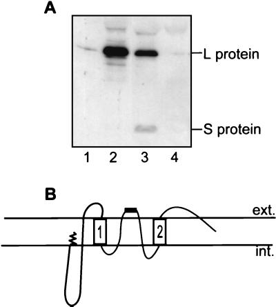
FIG. 4.
The loop region between TM1 and TM2 protrudes through the particle surface. (A) SVPs were immunoprecipitated with antibody to aa 10 to 29 (lane 1), aa 127 to 156 (lane 2), or aa 207 to 226 (S loop) (lane 3) and with a control antiserum (lane 4). Antibody prebound to protein G-Sepharose was incubated with SVPs, which were then disrupted with RIPA buffer. The pelleted immune complex, representing protein subunits captured via binding of antibody to external or exposed epitopes on whole SVPs, was separated by SDS-PAGE and detected by Western blotting with anti-pre-S and anti-S domain (aa 267 to 284) monoclonal antibodies. (B) Revised topology of L with the loop region between TM1 and TM2 being shown as membrane embedded and with part of the loop, containing the epitope of aa 207 to 226, shown as a black bar, being exposed to the lumen and ultimately to the particle surface. ext., exterior; int., interior.

