Figure 2.
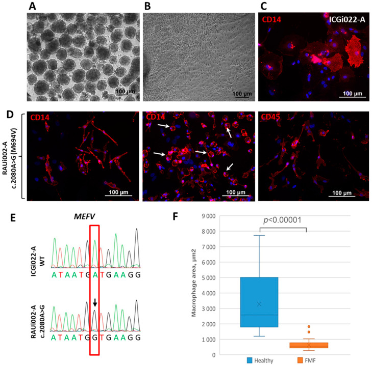
Differentiation of iPSCs into macrophages and characteristics of the resulting cells. (A) Morphology of embryoid bodies on the 4th day of differentiation of RAUi002-A iPSC line. (B) Morphology of spread out embryoid bodies on the 5th day after plating of the RAUi002-A line. (C) Immunofluorescence of CD14 on macrophages derived from control iPSCs line ICGi022-A. (D) Immunofluorescent of CD14 and CD45 on macrophages derived from RAUi002-A iPSC line. White arrows indicate rounded cells. Nuclei were stained with DAPI (blue signal). All scale bars: 100 μm. (E) Sanger sequencing confirmed the presence of c.2080A>G (M694V) mutation in macrophages derived from RAUi002-A iPSCs. The position of the detected polymorphism indicated with red box. The detected polymorphism is marked with arrow. (F) Comparison of average area of macrophages derived from FMF patient’s iPSCs and a healthy donor. n = 50, Mann–Whitney U test was used to assess statistical significance. p-value < 0.00001.

