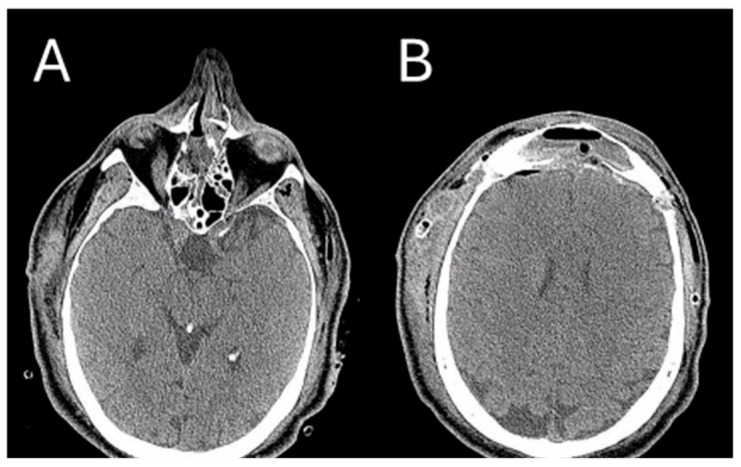
Figure 6.
Post-operative CT image of Patient 2. Two axial cuts of the post-operative CT scan are appreciatedat the same level as those shown in Figure 3 to demonstrate the outcomes of the surgical removal. (A) the absence of the lesion localized at the ethmoidal level (B) The absence of the lesion localized in the right frontal sinus.

