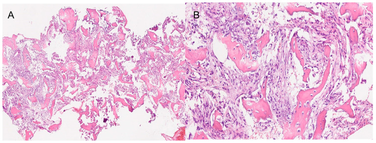
Figure 7.
The histopathological images show a section of the bones samples. (A) 4× magnification. (B) 20× magnification. Both images show the fibrous component with variable, medium to low cellularity. Osteoblastic rimming is observed in all trabeculae. The osseous component exhibits amorphous crystalline, calcific deposits. Segments of respiratory-type epithelium with areas of squamous metaplasia are also observed.

