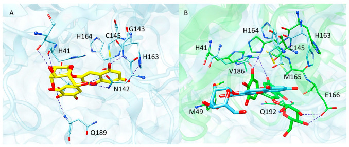
Figure 9.
(A) Docking pose of delphinidin-3-glucoside (yellow sticks) within Mpro active site. (B) Representative structure of the MD simulation performed on delphinidin-3-glucoside–Mpro complex (green) superimposed to the starting coordinates obtained from docking (cyan). The residues of the binding pocket are displayed as sticks, while blue dashed lines represent H-bond interactions.

