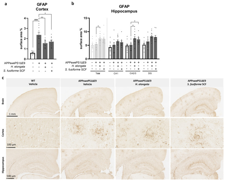
Figure 6.
H. elongata extract and S. fusiforme SCF extract reduce the elevated astrocytic marker GFAP in the cortex of APPswePS1ΔE9 mice but increase GFAP in the hippocampus. The quantification of the immunohistochemical staining of GFAP in the cortex (a) and hippocampus (b) of WT and APPswePS1ΔE9 mice are presented as relative surface area. Representative images of the GFAP staining are presented in (c). Data are represented as mean ± SEM (n = 10–11 per group, 3 cortical and 1 hippocampal image of 1 slide per animal). Differences between vehicle-treated APPswePS1ΔE9 and WT mice were analyzed with an unpaired t-test (a) or Mann–Whitney U test (b) (### p < 0.001); Treatment effects in APPswePS1ΔE9 mice were analyzed with a one-way ANOVA (with Dunnett’s multiple comparisons test) (a) or a Kruskal–Wallis test (with Dunn’s multiple comparisons test) (b) (* p < 0.05, ** p < 0.01).

