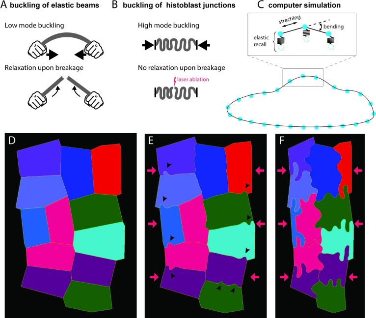
Fig 5. Computer simulation of buckling.
(A, B) Some features of histoblast buckling are incompatible with simple elastic beam buckling. (A, top) An elastic beam under compressive stress at the extremities buckles through low mode deformation. (A, bottom) The stored bending energy is released upon breaking the beam, which results in a recoil of the beam. (B, top) Histoblasts buckle at small wavelengths (high mode). (B, bottom) Upon laser dissection, junctions hardly relax. (C) Computer simulations of histoblast mechanics. Cell boundaries are represented as point masses (vertices) connected by springs (edges). The elastic energy of the boundary includes stretching and bending terms. Connection to the elastic environment is simulated with springs that pull vertices back to their position. (D-F) Snapshots of the computer simulation of an histoblast nest going through the morphological transition. In the initial configuration, cells are polygonal (D). Early on in the transition, isolated lobules appear along the cell–cell interfaces (arrowheads in E). Lobules have fully developed in the final state.

