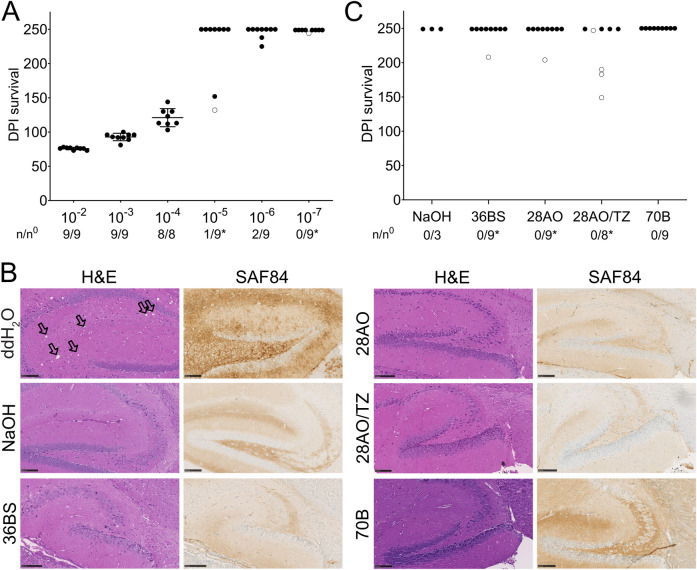
Fig 3. Mouse bioassay for the evaluation of the decontamination efficiency of the different formulations.
(A) End-point titration in tga20 mice inoculated with beads exposed to 10-fold serial dilutions of RML6 from 10−2 to 10−7. Data points are shown as the mean incubation time ± standard deviation (SD) of the mean for dilutions 10−2 to 10−4. n/n0: indicates the attack rate (number of mice developing a prion disease divided by the total number of inoculated mice). Open circles: individual mice that died of an intercurrent death unrelated to prion infection. (B) Histopathology of brain sections of the hippocampus from tga20 mice inoculated with RML6-exposed beads treated with either ddH2O, NaOH or the different formulations (as indicated in the Figure). Vacuolation along with PrPC and PrPSc deposition, serving as markers for the presence of prion disease, was observed upon visualization through H&E staining and imaging with an antibody targeting PrP (SAF84) in brain sections of mice inoculated with RML6-coated beads treated with ddH2O (see also S3 Fig). No vacuolation and PrPSc deposits were observed in the brains of mice inoculated with RML6-coated beads treated with NaOH or the different formulations. The SAF84 staining for 70B is more intense than for the other slides due to a stronger background signal of PrPC resulting from the use of a thicker slide that was necessary as the sectioning was affected by the presence of the beads in the brain. The use of a thicker slide also causes a more intense H&E staining for 70B. Coronal sections are presented for mice inoculated with RML6-coated beads treated with ddH2O, while sagittal sections are shown for all other mice. Vacuoles are indicated by black arrows. (Scale bars: 100 μm). (C) Survival of tga20 mice inoculated with RML6-exposed beads treated either with NaOH or the different formulations. None of the mice developed a prion disease until the end of the experiment.

