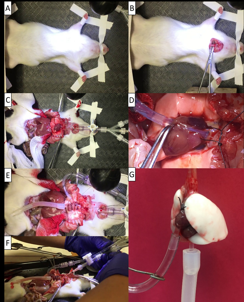
Figure 3: Isolation process of the rat lungs.

(A) The rat is securely taped down after anesthesia with the mouth taped as well to ensure minimal movement. (B) Proper isolation and placement of the forceps for performing the tracheostomy. (C) Spreading the rib cage to open the surgical area and minimize the risk of a broken rib puncturing the lungs. (D) Placement of the pulmonary artery cannula. (E) Placement of the pulmonary vein cannula following it being secured to the heart. (F) Removal of the trachea and heart-lung block from the rat. (G) The rat isolated lungs hanging prior to placement in the chamber.
