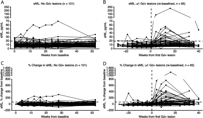
Figure 1. Timecourse of sNfL Levels According to Gd+ Lesions Status.
Individual subject sNfL levels over time among subjects without Gd+ lesions (absolute values A, percentage changes C) and with Gd+ lesions (absolute values B, percentage changes D). Horizontal dotted line in A and B represents 19.6 pg/mL change in sNfL from baseline. Horizontal dotted line in C and D represents the 68.8% change in sNfL from baseline. Vertical dotted line in B and D represents time of first detected Gd+ lesion (week 0). Gd+ = gadolinium-enhancing; sNfL = serum neurofilament light chain.

