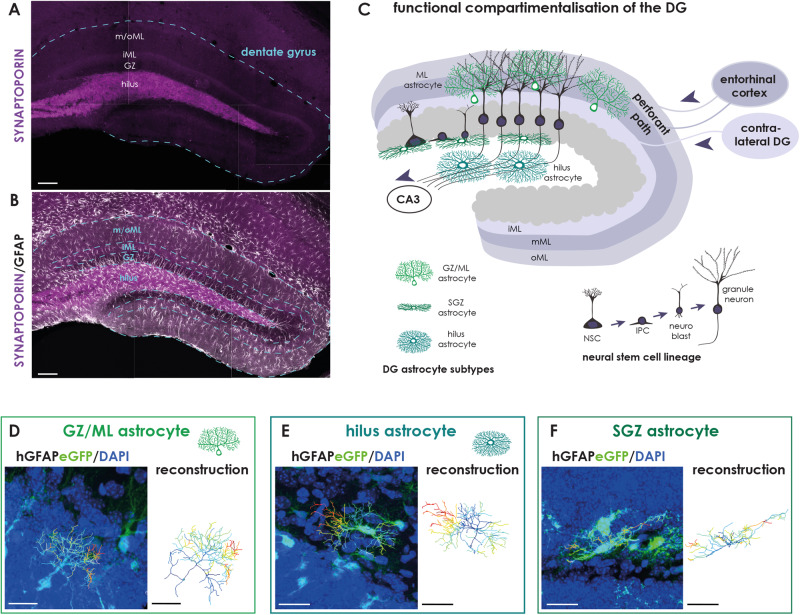
Fig. 1. Astrocyte subtypes in the adult DG are associated to distinct functional compartments.
A Confocal image of the adult DG (blue line) immunostained against SYNAPTOPORIN (magenta) illustrating the distinct layers (hilus, GZ granule zone, iML inner Molecular layer; m/oML medial and outer Molecular layer). B Astrocytes of the adult DG as revealed by GFAP immunostaining (white). C Schematic depicts the compartmentalization of the adult DG. Afferents from the entorhinal cortex form the medial and outer ML, while those from the contralateral DG form the inner ML. The neurogenic lineage (gray) consists of a neural stem cell (NSC) giving rise to actively proliferating intermediate precursor cells (IPCs), which generated neuroblasts (NBs). NBs subsequently mature to granule neurons. D–F Astrocytes reveal a subtype-specific morphology as determined by the expression of enhancedGFP (green) in brain slices of hGFAPeGFP transgenic animals (on the left). The eGFP signal was reconstructed in IMARIS (depicted on the right); different colors highlight branching order (dark blue=primary; light blue=secondary; turquoise=tertiary; green=quaternary; light green=quinary; yellow=septenary; orange=octonary; red = nonary). D Astrocytes of the upper DG compartments (ML and GZ) show a polarized morphology with one primary process emerging from the soma and fanning out into the ML with treetop-like branches. E Hilus astrocytes reveal a bushy morphology with several primary processes pointing in each direction. F SGZ astrocytes are smaller and stretch out one to two primary branches along the SGZ boarder. Scale bar = 100 μm (A, B) and 20 μm (D–F).

