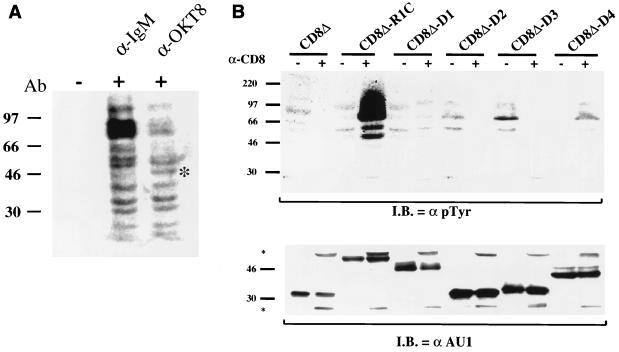
FIG. 5.
Induction of cellular tyrosine phosphorylation upon antibody stimulation. (A) Comparison of anti-IgM- and anti-CD8-stimulated CD8Δ-R1C cells. Cross-linking of the CD8Δ-R1C cell line with an anti-IgM or anti-CD8 (OKT8) antibody (Ab) induced tyrosine phosphorylation of cellular proteins as determined by Western analysis using an anti-pTyr antibody. Asterisk, 50-kDa phosphorylated protein seen in the CD8 antibody-stimulated, but not IgM antibody-stimulated, B cells. (B) Mutational analysis of CD8Δ-R1 chimeras for the induction of tyrosine phosphorylation upon antibody stimulation. Cells were incubated in the absence (−) or presence (+) of anti-CD8 antibody for 1 min at 37°C and immediately lysed. Cell extracts were subjected to gel electrophoresis, transferred to nitrocellulose, and reacted with an anti-pTyr antibody (top). Cell extracts were probed with an anti-AU1 antibody (bottom) to show comparative levels of chimeric proteins in these cells. Asterisks, heavy and light chains of the CD8 antibody used for stimulation. I.B., immunoblotting.

