Figure 4.
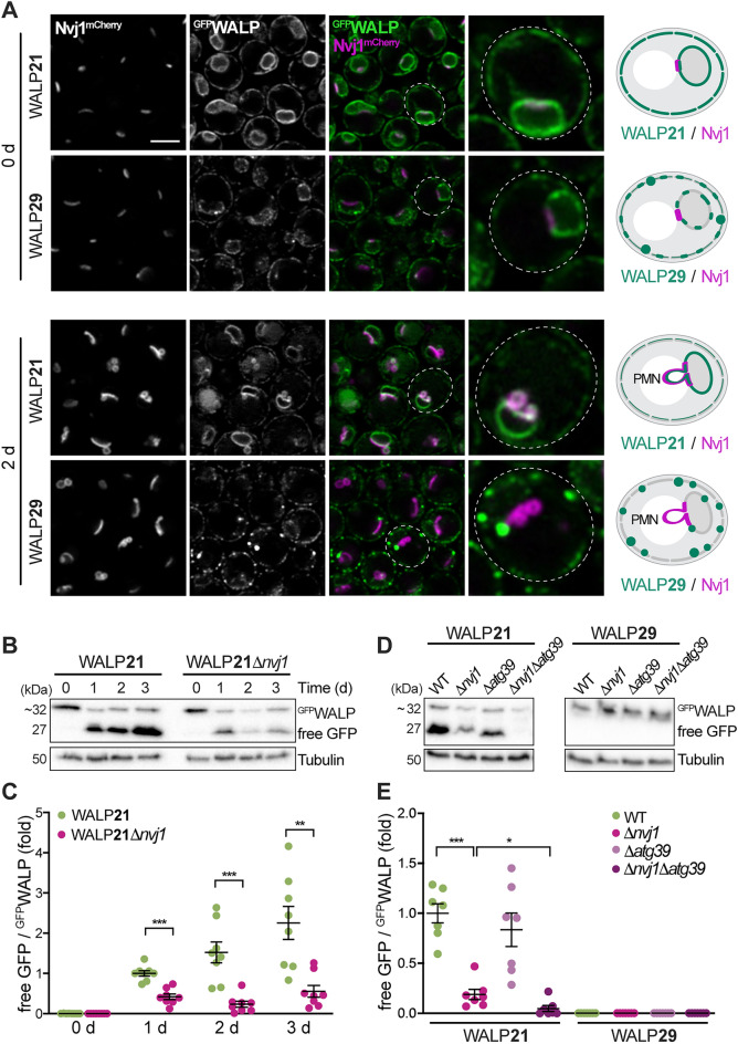
Reporters with short TMD are removed via piecemeal microautophagy of the nucleus. (A) Micrographs of exponentially growing and stationary cells endogenously expressing Nvj1mCherry and GFPWALP21 or GFPWALP29. Scale bar: 3 μm. Schematics of TMD length-dependent exclusion of WALP29 from the nucleus vacuole junction (NVJ) and piecemeal microautophagy of the nucleus (PMN). (B, C) Immunoblot analysis of total protein extracts from wild type (WT) and Δnvj1 cells expressing GFPWALP21 collected at indicated time points during chronological aging. A representative blot (B) and corresponding densitometric quantification of the ratio of free GFP to GFPWALP21 (C) are depicted. Blots were decorated with antibodies directed against GFP and tubulin as loading control. The free GFP/GFPWALP values are shown as fold of the ratio at day 1. Dot plots with mean ± s.e.m.; n = 8. (D, E) Immunoblot analysis of total protein extracts from WT, Δnvj1, Δatg39, and Δnvj1Δatg39 cells expressing GFPWALP21 or GFPWALP29 collected at day 2. Representative blots (D) as well as corresponding densitometric quantification of the ratio of free GFP to GFPWALP (E) are depicted. Blots were probed with antibodies directed against GFP and tubulin as loading control. The free GFP/GFPWALP values are shown as fold of the ratio of GFPWALP21 in WT. Dot plots with mean ± s.e.m.; n = 7. *p ≤ 0.05, **p ≤ 0.01, and ***p ≤ 0.001.

