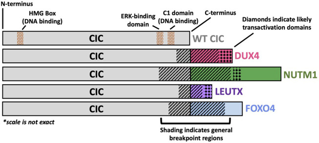
FIGURE 1.
Structural Schematic of CIC Fusion Oncoproteins. Structural schematic of CIC fusions highlighting the highly conserved wild-type (WT) CIC region with variability in the fusion break point and c-terminal binding partner. The CIC::DUX4 fusion oncoprotein has been shown to retain WT CIC DNA binding specificity through its HMG box and C1 domain but acquires transcriptional activating capacity via DUX4-mediated p300 recruitment to CIC binding sites. The C-terminal bindings partners, NUTM1, LEUTX, and FOXO4 have previously been reported to interact with p300/CBP.

