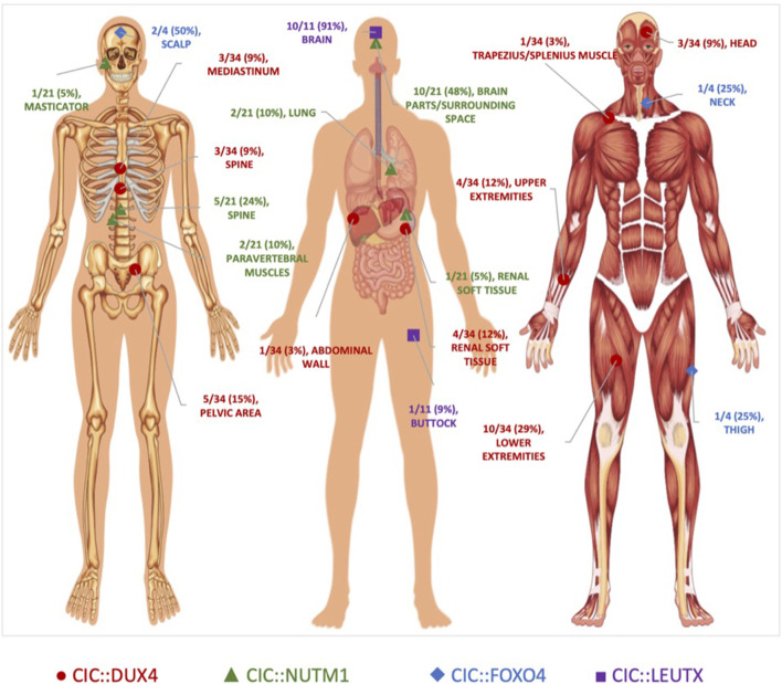
FIGURE 2.
Anatomical Localization of CIC-rearranged Primary Tumors. Primary tumor location of CIC-fusion positive cases reported in the literature. CIC::DUX4 (red circles) tumors localized primarily in the soft tissue. CIC::NUTM1 (green triangles) tumors were predominantly observed in the central nervous system. CIC::LEUTX (purple squares) tumors were confined mostly in the brain, while CIC::FOXO4 (blue diamonds) tumors were interspersed in the scalp, neck, and thigh. Original unlabeled anatomical image from Vecton/Shutterstock.com.

