Fig. 3.
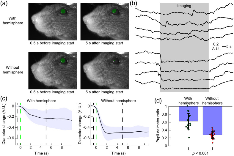
Aluminum hemisphere shields illumination and reduces pupil constriction during mesoscale imaging. (a) Images of the mouse face 0.5 s before and 5 s after widefield imaging acquisition begins. Green circles highlight the pupils. (b) Example 2-min pupil diameter time courses showing the off-to-on and on-to-off transition with (top) and without (bottom) hemisphere. The gray-shaded area indicates the period when widefield acquisition was performed. (c) Average pupil diameter changes upon beginning of widefield imaging (20 trials; traces show average ± standard deviation). Pupil diameter 1.5 to 0.5 s before imaging was defined as baseline for each trial. The green-dashed line indicates the start of widefield imaging, the black dashed lines indicate time points of the images shown in panel (a). (d) Comparison of the ratio of pupil size 2 to 3 s after start of imaging to pupil size 1.5 to 0.5 s before imaging. Significance was tested with a student’s test (Video 1, MP4, 5.93 MB [URL: https://doi.org/10.1117/1.NPh.11.3.034310.s1]).

