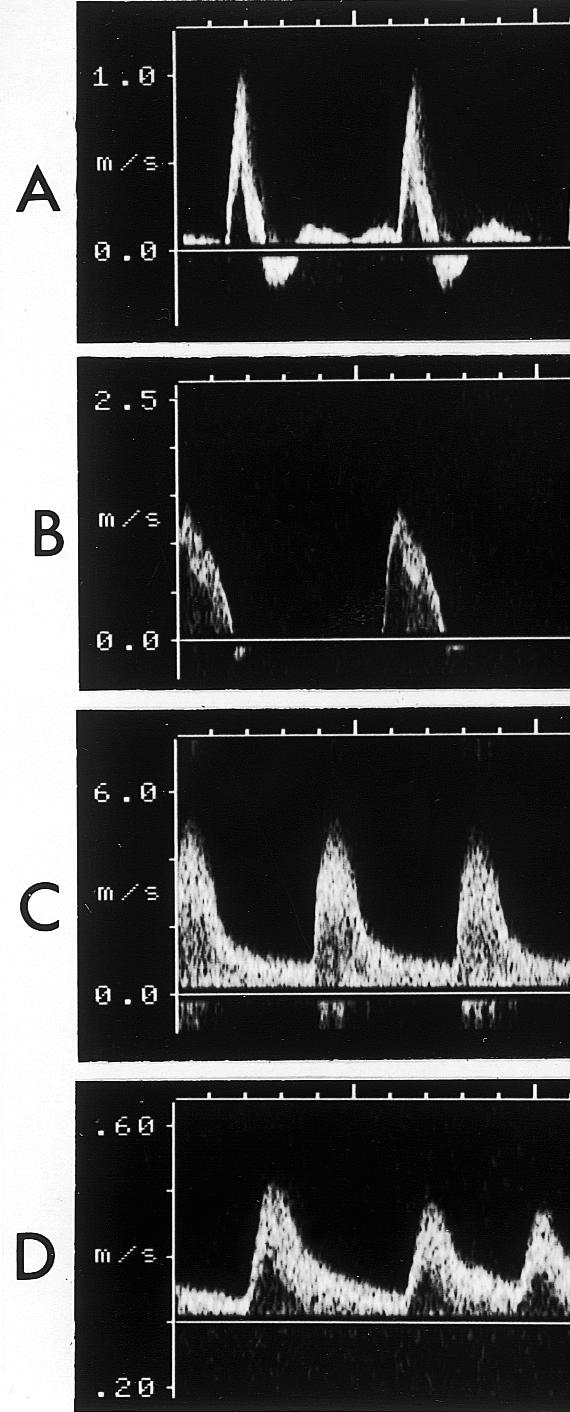
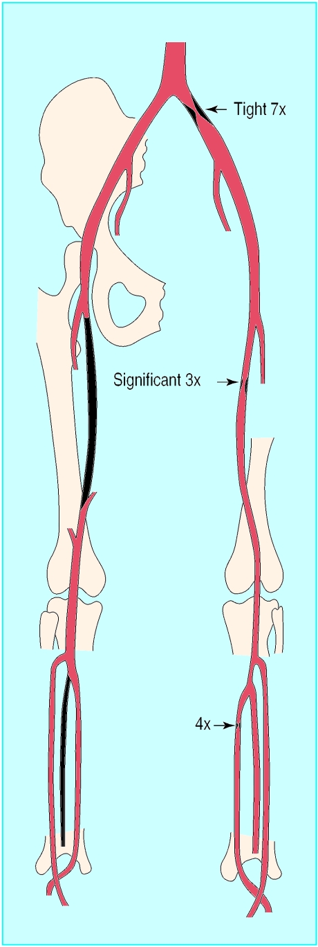
Figure.

Left: Doppler velocity waveforms: (a) triphasic waveform in normal artery; (b) biphasic waveform, with increased velocity, through a mild stenosis; (c) monophasic waveform, with greatly increased velocity, through tight stenosis; and (d) dampened monophasic waveform, with reduced velocity, recorded distal to tight stenosis. Right: Anatomical chart used to record position of stenoses, showing three stenoses with velocity increases of 7×, 4×, and 3× compared with adjacent unaffected arteries

