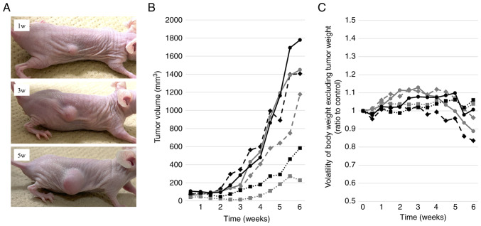
Figure 2.
Tumor progression and weight transition of the PDX model. PDX tissue pieces (size, ~3 mm) were transplanted into the backs of nude mice and tumor progression was monitored for 6 weeks. The tumor diameter and body weight were measured twice a week. (A) Representative images of the PDX model at 1, 3 and 5 weeks post-transplantation. Plots of (B) tumor volume progression and (C) body weight volatility excluding estimated tumor weight. The tumor weight was calculated using the following formula: Long diameter × (short diameter)2/2. PDX, patient-derived xenograft.

