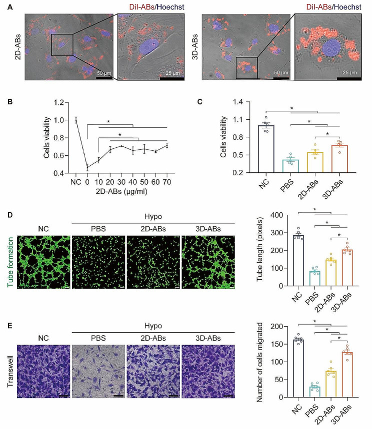
Fig. 2.
3D-ABs promoted the survival of HUVECs. (A) Uptake of 2D and 3D-ABs in HUVECs detected by confocal microscopy. (B) Proliferation of Hypo-HUVECs after 2D-ABs treatment measured by CCK-8 assays. (C) Proliferation of Hypo-HUVECs after 2D and 3D-ABs treatment measured by CCK-8 assays. (D) After treatment for 24 h, Hypo-HUVECs were divided into four groups and exposed to an in vitro angiogenesis (tube formation) experiment. The findings showed that the cells could be cultured for six hours. Scale bars, 100 μm. Quantification of tube length (pixels) among the four groups (n = 6). (E) After treatment for 24 h in each of the four groups, Hypo-HUVECs were subjected to cell migration experiments; the data shown here were obtained after 12 h of culture. Scale bars, 100 μm. Quantification and analysis of the number of cells migrating throughout a 12-hour period in each of the four groups (n = 6). SEM error bars are used. Significance (*): p value < 0.05; equal variances ANOVA with LSD post hoc analysis or unequal variances Dunnett’s T3 technique

