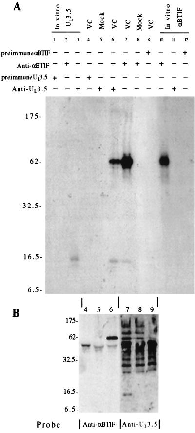
FIG. 2.
Coimmunoprecipitation of UL3.5 and αBTIF from virus-infected cells. (A) Cells were infected with BHV-1 at an MOI of 10 or were mock infected and labeled with [3H]leucine for 12 h beginning at 6 h pi. Proteins from BHV-1-infected (VC) and mock-infected (Mock) cells were precipitated with anti-UL3.5 antibodies (lanes 5 and 6) or anti-αBTIF antibodies (lanes 7 and 8). Proteins from BHV-1-infected cells were also precipitated by UL3.5 and αBTIF preimmune sera (lanes 4 and 9). The UL3.5 protein was synthesized in vitro in the presence of [3H]leucine and was immunoprecipitated with anti-UL3.5, anti-αBTIF, and UL3.5 preimmune sera (lanes 3, 2, and 1, respectively). [35S]Met- and [35S]Cys-labeled in vitro-synthesized αBTIF was immunoprecipitated with anti-αBTIF, anti-UL3.5, and αBTIF preimmune serum (lanes 10 to 12, respectively). The precipitated proteins were analyzed by SDS-PAGE on a 10 to 20% gradient gel and fluorographed. (B) One-fifth of samples 4 through 9 from panel A were separated by SDS-PAGE on a 13% gel. The proteins were transferred onto a nitrocellulose membrane and probed with either anti-UL3.5 or anti-αBTIF antibodies as indicated. The bound antibodies were detected by enhanced chemiluminescence.

