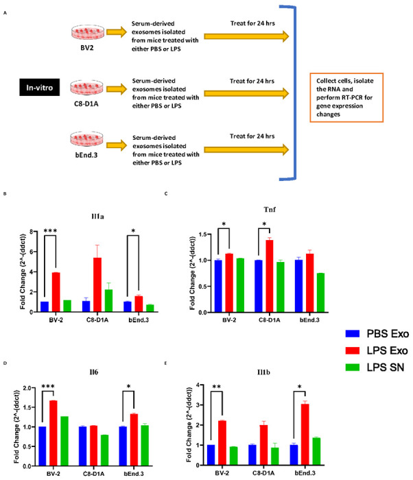
Figure 2.

(A) Scheme depicting the in vitro experimental design for investigating the effects of serum derived exosomes. (B-E) mRNA fold change levels of various proinflammatory genes in the cell lines BV-2 (microglia), C8-D1A (astrocytes) and bEnd.3 (brain endothelial cells) treated for 24 hours with 0.1 mg/ml of exosomes derived from the serum of the donor mice that were injected with PBS (PBS Exo), LPS (LPS Exo) or LPS derived exosome supernatant (LPS SN) for 24 hours, (B) Il1a (C) Tnf, (D) Il6, (E) Il1b (n= 3-4 male mice in each group, Data represent mean ± SEM; Two-way anova with Dunnett post-hoc test, *p<0.05, **p<0.01, ***p<0.001)
