
Figure 5. NADH blocks formation of the immunoproteasome.
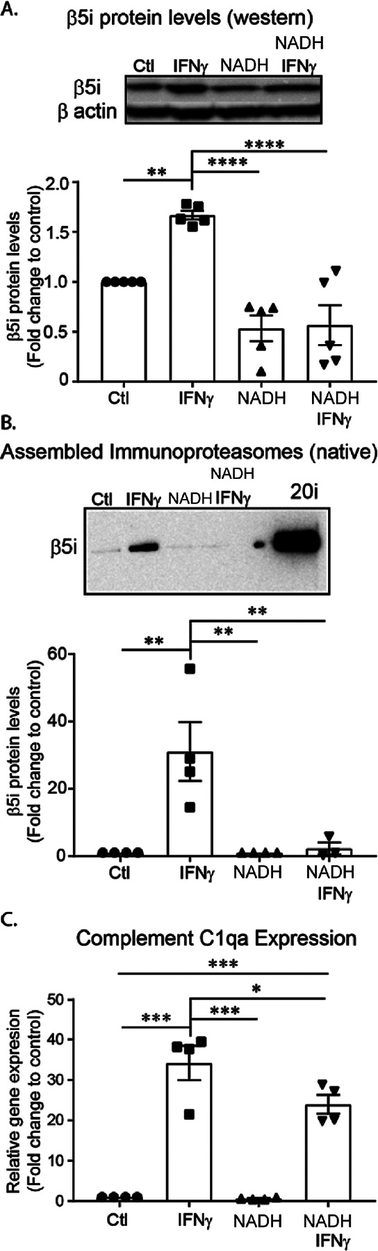
BV-2 cells were pre-treated with NADH for 24 hours, then treated with IFNγ for an additional 24 hours. Relative amounts of immunoproteasome protein levels were quantified. A. Western blot analysis revealed a significant difference between treatment groups ([F(3, 16) =19.04], p<.001, n=4). Post hoc analysis revealed that IFNγ increasesd total β5i protein levels compared to all groups (Control, p=.006; NAD, p<.001; NADH+IFNγ, p<.0001). Further, IFNγ did not significantly increase β5i protein levels in cells pre-treated with NADH (p=.997). B. Assembled immunoproteasomes (20i represents purified positive control) were measured using native gel electrophoresis. Analysis revealed that there was a significant difference between treatment groups ([F(3, 11) =9.845], p=.002, n=4). Post hoc analysis revealed that IFNγ increased the amount of assembled immunoproteasomes compared to all treatment groups (Control, p=.003; NADH, p=.004; NAD+IFNγ, p=.009). Interestingly, when cells are pre-treated with NADH, immunoproteasomes are not increased in response to IFNγ (p=.996). C. To determine if NADH treatment would successfully reduce complement activation in BV-2 cells, we pre-treated with NADH, then measured gene expression of complement activator C1qa. An ANOVA revealed a significant treatment effect ([F(3,12)=48.22, p<.001). Post hoc analysis revealed a significant increase of C1qa gene expression in response to IFNγ treatment (p<.001), an effect that was reduced by NADH pre-treatment (p=.049).