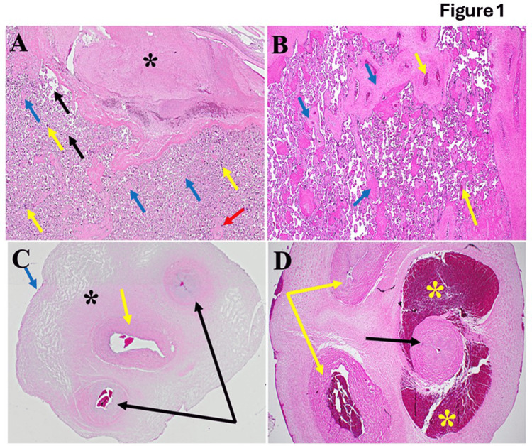
Figure 1. Architecture of placental tissues in normal and postmenopausal pregnancy women.
(A) Illustrating the typical architecture of placental tissues, the decidua is well-formed (black star). The intervillous space is clear, with few red blood cells (black arrow). Blue arrows point to the cytotrophoblast surrounded by syncytiotrophoblasts (yellow arrows). The red arrow points to villous blood vessels containing fetal blood. (B) shows diseased placental tissues; we observed significant disruption of the typical architecture of the decidua compared to Figure 1A (blue arrows). The yellow arrow indicates congested blood vessels. Additionally, scattered inflammatory cells and increased collagen deposition were noted. (C) The normal allantoic anatomy is illustrated by a single central artery (yellow arrow), paired veins (black arrows), and the black star, which indicates Wharton’s jelly. At the same time, the amniotic epithelium is marked by the blue arrow. On the other hand, this is evident in the fact that funisitis scatters diffusely (D). The artery (black arrow) is surrounded by severe congestion (yellow stars). Furthermore, inflammation of the veins' walls (phlebitis) and increased collagen deposition (yellow arrows) are noted.

