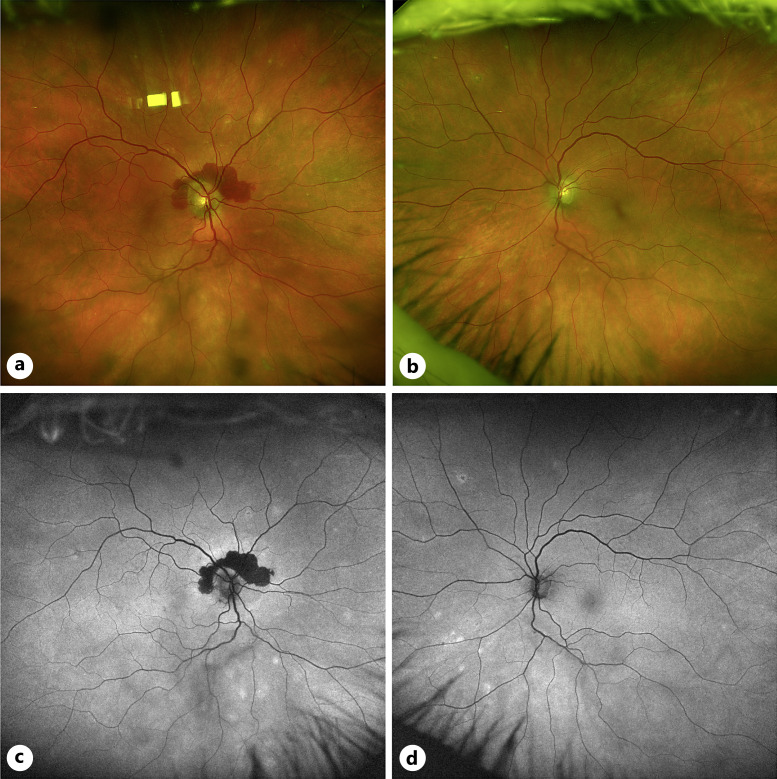
Fig. 1.
Fundus images of case 1. a Color fundus photo of the right eye with notable peripapillary hemorrhage and mid-peripheral RPE changes. b Color fundus photo of the left eye with mid-peripheral RPE changes. c Fundus autofluorescence imaging showing hypoautofluorescent peripapillary signal corresponding to the hemorrhage in (a) with hyperautofluorescent changes in the mid-periphery correspond with RPE changes in (a). d Fundus autofluorescence imaging showing hyperautofluorescent changes in the mid-periphery correspond with RPE changes in (b).

