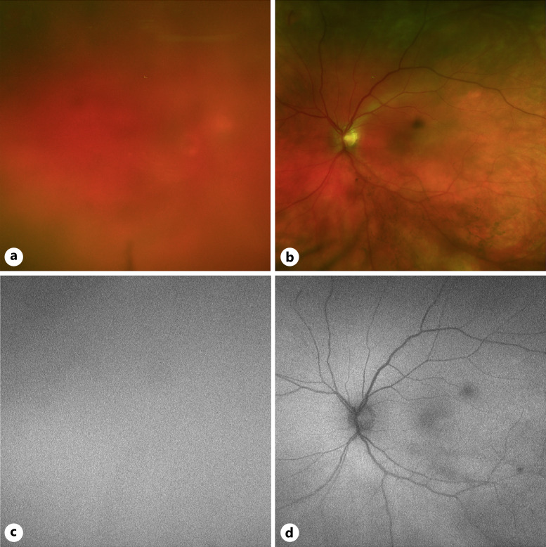
Fig. 6.
Fundus images of case 3. a Color fundus photo of the right eye was obstructed due to dense vitreous cell. b Color fundus photo of the left eye with vitreous opacities, blunted foveal reflex, and inferior periphery with reticular pigmentary changes. c Fundus autofluorescence imaging of the right eye yielded no diagnostic information due to vitreous cell. d Fundus autofluorescence imaging showing autofluorescent changes aligned with vitreous debris.

